Thoracoscopy
Thoracoscopy
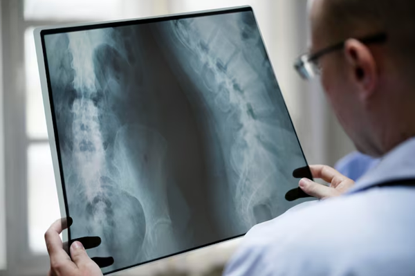
Thoracoscopy is an advanced minimally invasive procedure used to diagnose and treat diseases affecting the lungs, pleura, and chest cavity. It allows pulmonologists to directly examine the inside of the chest using a specialized instrument called a thoracoscope. This procedure is highly effective in identifying the cause of pleural effusion, lung infections, pleural diseases, tuberculosis, and certain types of lung cancer.
At Wadhwan Health Care Centre, thoracoscopy is performed using modern technology and advanced pulmonary expertise to provide accurate diagnosis and effective treatment for complex chest conditions. The procedure helps avoid large surgical incisions and enables faster recovery with minimal discomfort. With expert respiratory specialists and comprehensive pulmonary care, patients receive precise diagnosis and personalized treatment for various thoracic and respiratory diseases.
What Is Thoracoscopy?
Thoracoscopy is a minimally invasive medical procedure in which a thin instrument with a camera and light source is inserted through a small incision in the chest wall to examine the pleural cavity and lungs. The camera provides detailed real-time images of the lungs, pleura, diaphragm, and chest cavity structures.
During the procedure, tissue samples (biopsy) or fluid samples may be collected for laboratory analysis. Thoracoscopy is commonly used for diagnosing unexplained pleural effusion, pleural infections, tuberculosis, lung cancer, and inflammatory chest diseases.
In some cases, thoracoscopy can also be used as a therapeutic procedure to drain fluid, remove infected tissue, or perform pleurodesis to prevent recurrent fluid accumulation.
Why Is Thoracoscopy Performed?
Thoracoscopy is recommended when imaging tests or routine investigations cannot clearly identify the cause of chest abnormalities. It provides direct visualization and accurate tissue diagnosis.
Common reasons for thoracoscopy include:
- Pleural effusion (fluid around the lungs)
- Recurrent chest fluid accumulation
- Suspected lung or pleural cancer
- Tuberculosis involving the pleura
- Pleural infections or empyema
- Persistent chest pain
- Unexplained lung abnormalities
- Lung nodules or masses
- Interstitial lung diseases
- Diagnostic pleural biopsy
The procedure is highly valuable for both diagnosis and treatment of various thoracic conditions.
Conditions Diagnosed and Treated Through Thoracoscopy
Pleural Effusion
Thoracoscopy helps determine the exact cause of fluid accumulation around the lungs and allows fluid drainage when necessary.
Pleural Tuberculosis
It assists in diagnosing tuberculosis affecting the pleura by obtaining tissue samples for laboratory testing.
Lung Cancer and Pleural Cancer
Thoracoscopy allows direct examination of suspicious lesions and collection of biopsy samples for accurate cancer diagnosis and staging.
Empyema
Empyema is a collection of infected fluid or pus in the pleural cavity. Thoracoscopy helps remove infected material and improve lung expansion.
Recurrent Pleural Fluid
The procedure can perform pleurodesis, which prevents repeated fluid buildup around the lungs.
Interstitial and Inflammatory Lung Diseases
Thoracoscopic biopsy may help diagnose complex inflammatory and fibrotic lung disorders.
Types of Thoracoscopy
Medical Thoracoscopy
Performed by pulmonologists under local anesthesia and sedation, primarily for diagnosis and pleural procedures.
Video-Assisted Thoracoscopic Surgery (VATS)
A more advanced surgical thoracoscopy performed under general anesthesia for major thoracic procedures.
Thoracoscopy Procedure
Before the Procedure
Patients may be advised to:
- Avoid eating or drinking for several hours before the procedure
- Inform the doctor about medications and allergies
- Stop blood-thinning medications if recommended
- Undergo blood tests and imaging studies before the procedure
During the Procedure
- The patient receives local anesthesia, sedation, or general anesthesia depending on the procedure type.
- A small incision is made in the chest wall.
- The thoracoscope is inserted into the pleural cavity.
- The doctor examines the lungs and pleura using real-time imaging.
- Tissue or fluid samples may be collected for biopsy.
- Therapeutic procedures such as fluid drainage or pleurodesis may be performed if required.
- A chest tube may be temporarily placed for drainage after the procedure.
The procedure duration may vary from 30 minutes to over an hour depending on the condition being treated.
Benefits of Thoracoscopy
Minimally Invasive Procedure
Thoracoscopy requires only small incisions, reducing pain and recovery time.
Accurate Diagnosis
Direct visualization and biopsy improve diagnostic accuracy for chest diseases.
Faster Recovery
Patients recover more quickly compared to open chest surgery.
Reduced Hospital Stay
Most patients require shorter hospitalization periods.
Therapeutic Advantages
The procedure allows both diagnosis and treatment during the same session.
Better Disease Management
Early and accurate diagnosis helps guide effective treatment planning.
Advantages Over Open Surgery
Compared to traditional open chest surgery, thoracoscopy offers:
- Smaller incisions
- Less pain and discomfort
- Reduced bleeding
- Lower infection risk
- Faster healing
- Minimal scarring
- Shorter recovery period
Recovery After Thoracoscopy
Recovery depends on the type and complexity of the procedure. Most patients experience mild chest discomfort for a few days.
Patients are usually advised to:
- Rest adequately after the procedure
- Avoid strenuous physical activity temporarily
- Follow medication and chest tube care instructions carefully
- Attend regular follow-up visits for biopsy results and recovery monitoring
Most patients gradually return to normal activities within a short period.
Risks and Complications
Thoracoscopy is generally safe when performed by experienced specialists. However, possible risks may include:
- Mild bleeding
- Infection
- Temporary chest pain
- Air leakage from the lungs
- Fever
- Breathing discomfort
- Rare complications related to anesthesia
Advanced monitoring and skilled pulmonary care significantly reduce complication risks.
Why Choose Wadhwan Health Care Centre for Thoracoscopy?
- Experienced pulmonology and thoracic care specialists
- Advanced thoracoscopy and respiratory diagnostic facilities
- Expertise in pleural diseases and lung disorders
- Minimally invasive pulmonary procedures
- Personalized patient-focused treatment
- Comprehensive chest disease management
- Accurate biopsy and pathology support
- Modern respiratory care infrastructure
The center is dedicated to delivering advanced pulmonary and thoracic care with precision, safety, and compassionate medical support.
Frequently Asked Questions (FAQs)
1. Is thoracoscopy painful?
Thoracoscopy is usually performed under anesthesia or sedation, so patients experience minimal pain during the procedure.
2. Why is thoracoscopy performed?
It is used to diagnose and treat pleural effusion, lung infections, tuberculosis, pleural diseases, and lung cancer.
3. How long does thoracoscopy take?
The procedure may take between 30 minutes and 1 hour depending on the complexity of the condition.
4. Is hospitalization required after thoracoscopy?
Yes, some patients may require short hospitalization for observation and chest tube management.
5. What is pleural effusion?
Pleural effusion is the accumulation of excess fluid around the lungs, causing breathing difficulty and chest discomfort.
6. Can thoracoscopy diagnose cancer?
Yes, thoracoscopy allows direct biopsy of suspicious lung or pleural lesions for accurate cancer diagnosis.
7. How long is recovery after thoracoscopy?
Recovery time varies, but most patients recover faster compared to traditional open chest surgery.
8. Is thoracoscopy safer than open surgery?
Yes, thoracoscopy is minimally invasive and generally associated with lower risks, less pain, and quicker recovery.